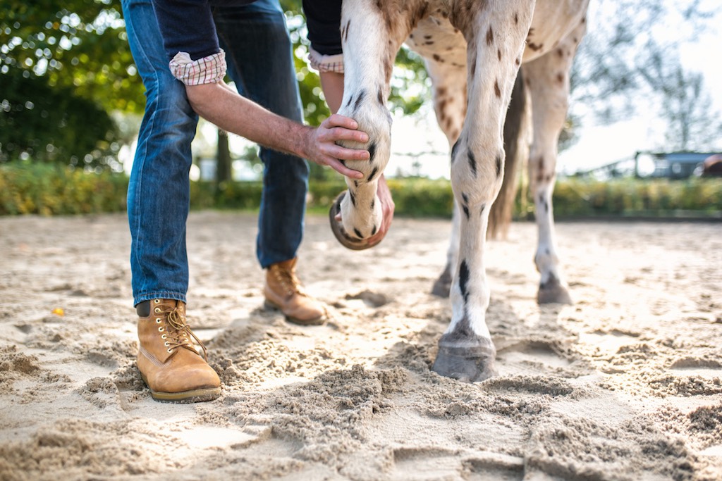
If you have horses, you will almost certainly discover a strange lump or bump on his leg one day. But should you rush to the phone and call the vet, apply ice or a cold hose, or simply take a ‘wait and see’ approach? A bit of investigation will help you make an initial assessment and determine if it’s a worrisome threat to your horse’s long-term soundness that needs immediate veterinary attention, or simply a blemish to be monitored. (If you are at all worried, or for any lump that feels warm to the touch, it’s best to call the vet).
Capped hock or elbow
Looks like: A swelling the size of a golf ball at the point of your horse’s hock or elbow. Your horse is sound.
Feels like: A soft lump with no heat that your horse doesn’t mind you handling.
Causes: Fluid accumulating in the thick walled bursa of the joint, as a result of repetitive concussion. Capped hocks are usually the result of a horse repeatedly kicking his stable wall or the side of the truck or float. A capped elbow is usually a response to pressure on the foreleg from the horse lying on a hard stable floor or from his shoe while his leg is tucked underneath him.
What to do: Usually a cosmetic problem rather than one that will affect soundness, but sometimes it will be large enough to cause mechanical lameness. The blemish is usually permanent, but if you address it early you can reduce or even reverse it. Call your vet as soon as you notice the swelling, as treatment in the first 24 hours has the best chance of preventing a firm and permanent lump forming. The vet may install a drain to remove the accumulated fluid, and steroid injections can reduce the swelling. Pressure bandages – though tricky to apply – can also help reduce the size of the bursa. Once the initial problem is dealt with, you’ll need to take steps to prevent a recurrence, such as putting rubber mats in your stable or truck to minimise the kicking, or putting a rubber ‘sausage’ boot around the fetlock to prevent pressure on the elbow.
Prognosis: If treated quickly, a capped hock or elbow can disappear completely, but once the lump firms up there’s a good chance it’s there to stay.
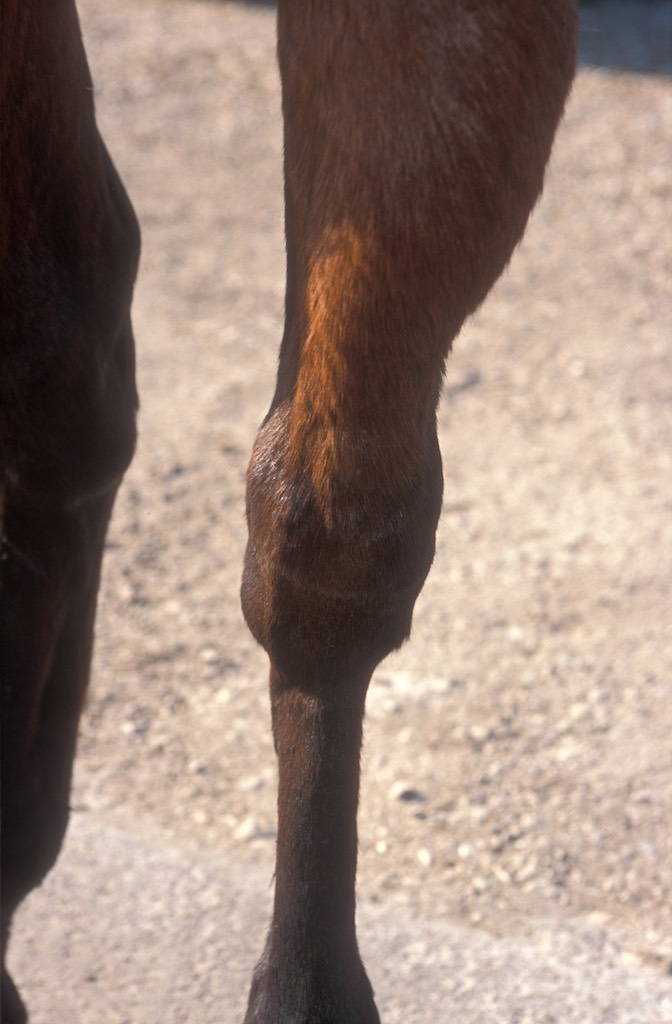
Bowed tendon

Looks like: A 5- to 7cm-long bulge running vertically down the back of your horse’s lower leg. From the side, it has a distinct ‘bow’. Your horse will be anywhere from three-legged lame to slightly off, and he won’t like you touching the area.
Feels like: A new bow will be soft and warmer than the rest of the leg, while an older injury is fairly firm and cold to touch.
Causes: Strain and tear of the superficial or the deep flexor tendon, that may also involve the suspensory or check ligament. Caused by trauma, repetitive stress or muscle fatigue from intense exercise. The bow occurs when broken tendon fibres leak blood and fill the torn area, creating a build-up of pressure that can further damage the surrounding tissue. Some conformation defects, such as long, weak pasterns, and poor or irregular shoeing, can also put horses at risk of bowed tendons.
What to do: A new bow is an emergency. Call your vet, tell him what you suspect, and follow his instructions. Your vet may want to ultrasound the leg straight away to determine the extent of the damage and administer anti-inflammatory drugs such as ’bute to make your horse more comfortable. He may also recommend icing the leg as often as possible during the first 24 hours. Your horse will likely be on box rest for a few weeks at least, then controlled turnout (in a small, flat paddock for a few hours at a time). After that, prepare yourself for anything between six weeks to 12 months of restricted work, starting with walking in-hand. And you’ll want your vet and your farrier to discuss the horse’s potential need for specialised trimming and shoeing to reduce the stress on damaged tissue.
Prognosis: A horse with a bowed tendon has a reasonably good chance of full recovery, depending on the severity of the injury. The scar tissue that will eventually fill the tear is not as elastic as the original tendon fibre, which means the leg will be prone to re-injury, especially if the horse is competing at a high-stress sport such as jumping or racing.
Ringbone
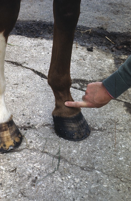
Looks like: Lumps around or above the coronary band. Your horse will be lame, especially on turns, and may have been off for some time, though the lameness might come and go depending on his activity level. He may also point the affected hoof, resting only the toe on the ground.
Feels like: Hard bony lumps that may feel warm and swollen to the touch, or hard and cold.
Causes: Ringbone is basically arthritis and degenerative joint disease in the coffin or pastern joint. The lumps are the bony growths formed in response to cartilage breakdown and inflammation. Trauma, particularly fracture of the coffin bone, can lead to the development of ringbone, as can poor conformation, and bad hoof trimming. Jumpers, polo ponies and other horses who compete in sports that involve quick, hard stops and fast turns are at higher risk for developing the problem.
What to do: Unfortunately, by the time you can see or feel the lumps, the condition is already at an advanced stage. Your vet can confirm the diagnosis with x-rays, which will also serve as a baseline to detect further changes. You can’t reverse the damage, but you can control the pain with medication and corrective shoeing. High ringbone, which affects the front of the pastern, is less debilitating than low ringbone, and a combination of rest, joint injections, anti-inflammatory medication and corrective shoeing can slow its progress.
Prognosis: Horses with high ringbone can typically continue in light work for months and even years if the condition is caught early and managed wisely. In rare cases, the pastern joint can fuse (or this can be done surgically) to end all motion and pain, but the horse may still not return to full soundness. Low ringbone means an end to any athletic career and the progressive retirement of pleasure horses.
Bog spavin
Looks like: A localised swelling on the front of the hock. Your horse is sound and doesn’t mind you touching the area.
Feels like: A soft lump that is no warmer than the rest of the hock.
Causes: Stress to the joint called the ‘tibiotarsal’, which is responsible for most of the movement of the hock. It can be acute stress, or a build-up, and this has caused the joint capsule to produce extra synovial fluid which collects under the skin, forming the spavin. A horse’s joint becomes ‘boggy’ for a number of reasons, including a kick or other trauma to the joint, hoof imbalance, poor conformation or hard use. It’s a common sign of some joint degeneration. But it may not affect soundness, or even lead to arthritic pain.
What to do: If your horse develops bog spavin as a result of a trauma, such as a kick, call the vet immediately. You’ll want to cold-hose it several times a day, for 20 minutes a time. The vet may also administer anti-inflammatory drugs such as ’bute. If there is no evidence of trauma, you will need to investigate why the spavin developed. Hoof balances or conformation faults can contribute to long-term soundness problems, so talk to your farrier about the possibility your horse will benefit from a different approach to trimming. You may also need to adjust your horse’s training to reduce the strain on his hocks.
Prognosis: Bog spavin alone isn’t career-ending, and one caused by a single trauma will disappear in a few weeks. But bog spavins that linger can be associated with a larger problem that will probably limit your horse’s athletic endeavours.
Windgalls

Looks like: Flat, round lumps about the size of a 50c piece, or larger, on and around the fetlock. Your horse isn’t lame and the lumps aren’t painful.
Feels like: Soft, puffy lumps that don’t emit any heat.
Causes: Windgalls are protrusions of the joint capsule and nearby tendon sheath, and typically appear after intense training sessions when stress on the joint triggers excess production of synovial fluid.
What to do: Bandaging and massage can reduce the size, but they will refill in a few hours. There’s no need for the vet, but you may want to discuss your horse’s trimming with your farrier, as hoof imbalance can be a contributing factor.
Prognosis: As long as there are no other issues, a windgall is simply a sign of high milage.
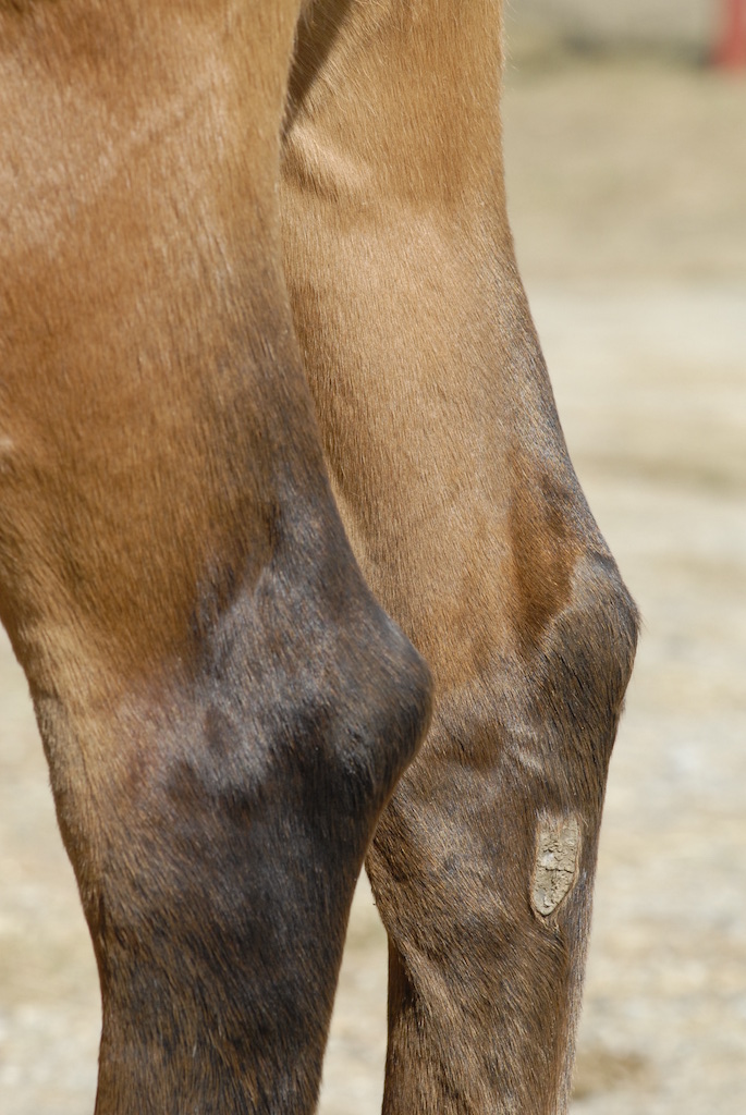
Splint

Looks like: A bony lump, from as small as a pea to as large as an egg, on the inside or the outside of a lower leg. If it’s a new lump, your horse may be slightly lame at the trot, and may flinch when you touch the area.
Feels like: A new splint may be hot to touch and somewhat soft at the edges. An older splint has hard, defined edges and is the same temperature as the rest of the leg.
Causes: Splints form when the ligament and the tissue that attaches the splint bone to the nearby cannon bone are stressed and tear. This produces a new bone, which creates a hard lump. The most common cause is work requiring tight circles or sharp turns, but splints can simply happen in the paddock as well, perhaps as a result of a knock. Faulty conformation, hoof imbalance and trauma to the area can also cause a horse to develop a splint, and they are most common in young horses just starting work. Correct diagnosis is important, as what looks like a common splint can be a fracture of the splint bone, which is more serious.
What to do: A fresh splint will benefit from 20 minutes’ cold-hosing, several times a day, to reduce the immediate inflammation. If your horse is lame, call the vet. If the bone is not fractured, any lameness should subside in a few days as the swelling goes down and the lump hardens. If it doesn’t, call the vet.
Prognosis: Splints are very common, and although the blemish can be visible for years, they only rarely pose a threat to soundness. Fractured splints may need surgery.
Thoroughpin

Looks like: Localised swelling on either the inside (medial) or the outside (lateral) face of the hock, just above the joint. Your horse is not lame and doesn’t react when you touch the lump.
Feels like: A soft, fluid-filled lump, that’s not hot, and can be pushed from one side of the leg to the other through the hollow just above the hock joint.
Causes: Thoroughpins are a swelling of the sac that holds lubricating fluid around the deep digital flexor tendon, just above the point of the hock. Most often, they are caused by your horse being asked to perform beyond his fitness level, though poor conformation and hoof imbalance are also factors.
What to do: Reduce your horse’s workload to match his fitness level, and keep a close eye on the size of the lump. Call the vet if the horse becomes lame (in which case, there are probably other issues at work). Thoroughpins usually reduce over time, but may never vanish. If the cosmetic appearance is important (as in a show horse), your vet may recommend injecting a steroid to reduce the inflammation, or withdrawing fluid.
Prognosis: Thoroughpins themselves won’t cause lamenesss, but until the stress on the hock is reduced, the lump will persist and other problems may develop. Sometimes disease of the large hock joint is misdiagnosed as thoroughpin.
- This article was first published in the June 2013 issue of NZ Horse & Pony